Year 2025 / Volume 36 / Number 2

Clinical Cases

Ostechondroma in the distal phalanx of a pediatric patient: a case report

Rev. Esp. Pod. 2025; 36(2): 139-142 / DOI: 10.20986/revesppod.2025.1751/2025

Alberto Rayo Martín, Rafael Rayo Martín, Rafael Rayo Rosado, Sandra Sánchez-Morilla, Raquel García de la Peña, Ana M.ª Rayo Pérez


ABSTRACT

Osteochondroma is a benign cartilage-derived tumor that, while common in the appendicular skeleton, is exceptionally rare in the distal phalanges of pediatric patients. Its clinical presentation may range from incidental findings to symptoms such as pain, local deformity, functional limitation, or recurrent infections, particularly when it affects exposed regions like the toes. These clinical features, combined with its unusual location, can complicate the differential diagnosis with entities such as exostoses, granulomas, or subungual infections, underscoring the importance of a thorough clinical and radiological assessment. We report the case of a 3-year-old girl presenting with a painful distal tumor in the fourth toe of the left foot, with a long-standing evolution and recurrent infectious episodes. Following comprehensive clinical evaluation and imaging studies, complete surgical excision was performed using Syme’s technique, with subsequent histopathological confirmation of osteochondroma. This case highlights the relevance of considering osteochondroma among less common differential diagnoses and emphasizes the effectiveness of surgical treatment in terms of safety, symptom control, and functional recovery. Moreover, early intervention not only ensures a definitive resolution but also prevents future complications, ultimately improving the patient’s quality of life.


RESUMEN

El osteocondroma es un tumor benigno de origen cartilaginoso que, aunque frecuente en el esqueleto apendicular, se presenta de manera excepcional en las falanges distales de pacientes pediátricos. Su expresión clínica puede variar desde un hallazgo incidental hasta síntomas como dolor, deformidad local, limitación funcional o infecciones recurrentes, especialmente en zonas expuestas como los dedos del pie. Estas características clínicas, sumadas a su rareza en dichas localizaciones, pueden dificultar el diagnóstico diferencial con otras entidades como exostosis, granulomas o infecciones subungueales, lo que subraya la importancia de un abordaje clínico y radiológico cuidadoso. Se expone el caso de una niña de 3 años con una lesión tumoral distal en el cuarto dedo del pie izquierdo, dolorosa, de evolución prolongada y asociada a episodios repetidos de infección. Tras la evaluación clínica y la realización de estudios de imagen, se procedió a la resección quirúrgica completa mediante técnica de Syme, confirmándose posteriormente el diagnóstico de osteocondroma a través del análisis histopatológico. Este caso ilustra la relevancia de considerar el osteocondroma en diagnósticos diferenciales poco habituales y destaca la eficacia del tratamiento quirúrgico en términos de seguridad, control sintomático y mejora funcional. Además, la intervención temprana favorece la resolución definitiva y previene complicaciones futuras, optimizando la calidad de vida del paciente pediátrico.


Complete Article


doi.org/10.20986/revesppod.2025.1751/2025
CLINICAL CASE

Ostechondroma in the distal phalanx of a pediatric patient: a case report

Osteocondroma en la falange distal de un paciente pediátrico: reporte de caso

Alberto Rayo Martín1
Rafael Rayo Martín2
Rafael Rayo Rosado2
Sandra Sánchez-Morilla3
Raquel García de la Peña2
Ana M.ª Rayo Pérez2

1Clínica Rayo. Arahal, Sevilla, España
2Departamento de Podología. Universidad de Sevilla, España
3Departamento de Enfermería y Podología. Universidad de Málaga, España

Abstract
Osteochondroma is a benign cartilage-derived tumor that, while common in the appendicular skeleton, is exceptionally rare in the distal phalanges of pediatric patients. Its clinical presentation may range from incidental findings to symptoms such as pain, local deformity, functional limitation, or recurrent infections, particularly when it affects exposed regions like the toes. These clinical features, combined with its unusual location, can complicate the differential diagnosis with entities such as exostoses, granulomas, or subungual infections, underscoring the importance of a thorough clinical and radiological assessment. We report the case of a 3-year-old girl presenting with a painful distal tumor in the fourth toe of the left foot, with a long-standing evolution and recurrent infectious episodes. Following comprehensive clinical evaluation and imaging studies, complete surgical excision was performed using Syme’s technique, with subsequent histopathological confirmation of osteochondroma. This case highlights the relevance of considering osteochondroma among less common differential diagnoses and emphasizes the effectiveness of surgical treatment in terms of safety, symptom control, and functional recovery. Moreover, early intervention not only ensures a definitive resolution but also prevents future complications, ultimately improving the patient’s quality of life.

Keywords: Osteochondroma, pediatric, bone pathology, benign tumor, surgical treatment, foot, toe, distal phalanx

Resumen
El osteocondroma es un tumor benigno de origen cartilaginoso que, aunque frecuente en el esqueleto apendicular, se presenta de manera excepcional en las falanges distales de pacientes pediátricos. Su expresión clínica puede variar desde un hallazgo incidental hasta síntomas como dolor, deformidad local, limitación funcional o infecciones recurrentes, especialmente en zonas expuestas como los dedos del pie. Estas características clínicas, sumadas a su rareza en dichas localizaciones, pueden dificultar el diagnóstico diferencial con otras entidades como exostosis, granulomas o infecciones subungueales, lo que subraya la importancia de un abordaje clínico y radiológico cuidadoso. Se expone el caso de una niña de 3 años con una lesión tumoral distal en el cuarto dedo del pie izquierdo, dolorosa, de evolución prolongada y asociada a episodios repetidos de infección. Tras la evaluación clínica y la realización de estudios de imagen, se procedió a la resección quirúrgica completa mediante técnica de Syme, confirmándose posteriormente el diagnóstico de osteocondroma a través del análisis histopatológico. Este caso ilustra la relevancia de considerar el osteocondroma en diagnósticos diferenciales poco habituales y destaca la eficacia del tratamiento quirúrgico en términos de seguridad, control sintomático y mejora funcional. Además, la intervención temprana favorece la resolución definitiva y previene complicaciones futuras, optimizando la calidad de vida del paciente pediátrico.

Palabras clave: Osteocondroma, pediatría, patología ósea, tumor benigno, tratamiento quirúrgico, pie, dedo, falange distal

Corresponding author
Alberto Rayo Martín
clinicarayo@gmail.com

Received: 23-07-2025
Accepted: 25-09-2025

Introduction

Osteochondromas are benign lesions characterized by hypertrophy of cartilaginous tissue. They are among the most frequent benign tumors, accounting for 10 % to 15 % of osteoarticular tumors(1,2). These cartilaginous tumors originate from a fragment detached from the epiphyseal cartilage, which determines their location. They generally develop after local trauma, although they may also appear without apparent cause(2,3,4).
They usually present as solitary lesions but may also show in multiple forms. They have been reported in nearly all bones of the skeleton, most frequently in the metaphyseal region of long bones(1,2,5).
Osteochondromas appear most often before the age of 20 years, and their discovery is usually incidental. When symptoms are present, these include deformity of the affected area, limitation of movement, and the presence of a palpable mass. Pain is typically related to compression of adjacent structures, as well as rubbing or friction against external objects(6,7). Diagnosis is primarily clinical but should be confirmed by imaging tests. Radiography is the method of choice, and osteochondromas are characterized by a bony protrusion with medullary continuity between the osteochondroma and the underlying bone. Other imaging modalities, such as ultrasound or magnetic resonance imaging, may also be used(5,6,8).
Surgery is indicated in cases with painful symptoms or when malignancy is suspected, such as in lesions with rapid growth or significant thickening of the cartilaginous cap(3,7,8).Among the most widely used techniques is the Mercado technique, which allows complete surgical resection of the tumor, including the cartilaginous cap and a small fragment of the underlying bone. This type of surgery requires full exposure of the tumor, which implies a wide approach for safe resection. It is often associated with moderate to severe postoperative pain due to greater tissue manipulation(8,9,10).
The aim of this work is to present, through a case report, the surgical approach to an osteochondroma in the distal phalanx in a pediatric patient. In this article, the term osteochondroma will be used consistently to refer to the lesion, with its synonymy and differences with chondroma explained in the introduction to avoid terminological ambiguities.

Case report

a 3-year-old female patient, with no relevant medical history, presented with a 15-month history of lesion on the distal region of the fourth toe of the left foot. The lesion caused pain and recurrent infections.
The case was initially evaluated by the Pediatrics service, which subsequently referred the patient to a podiatry unit specialized in surgery within a hospital setting, as the intervention would require general anesthesia and specific surgical management.
At the first consultation (June 2023), the parents reported difficulty in shoe use due to pain and recurrent infectious episodes, which temporarily improved with antibiotic treatment.
On clinical examination, the patient was in good general condition, with no motor dysfunction in the lower limbs. A distal tumoral lesion was observed on the 4th toe of the left foot, compromising the fingertip and producing nail dystrophy due to elevation of the nail plate (Figure 1). The lesion had well-demarcated borders, erythematous skin on its surface, and a small ulcerated area covered by a crust. Pain was elicited on palpation.

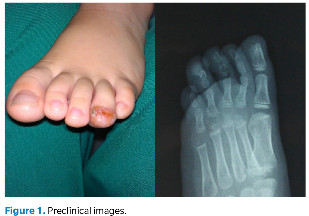
Plain radiography (anteroposterior and oblique views) showed a well-demarcated, bone-dense image originating in continuity with the distal phalanx of the 4th toe, consistent with the diagnosis of subungual osteochondroma.
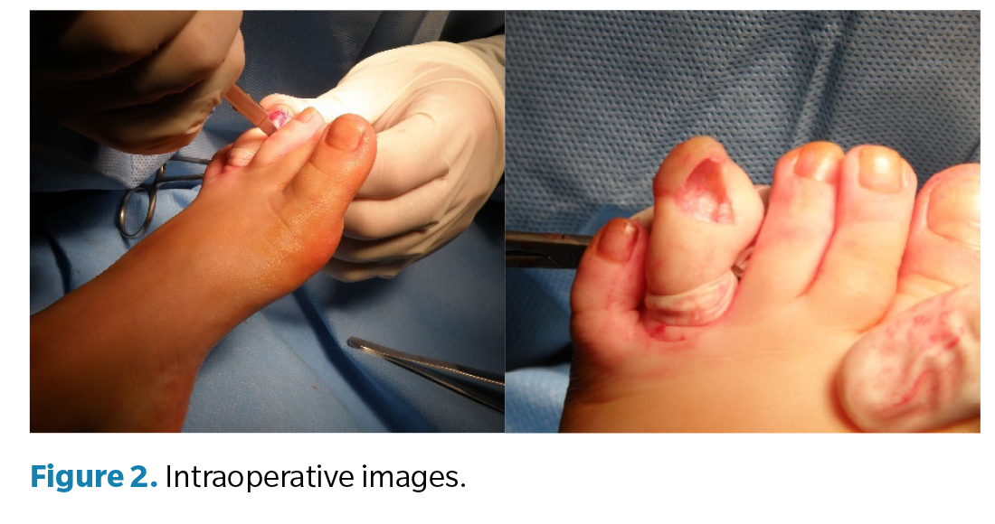
Given the location, functional compromise, and recurrence of infections, a block resection was performed using the Syme technique with complete removal of the distal phalanx, under general anesthesia, in August 2023. The choice of this technique, more radical than the conservative curettage recommended in the literature, was justified by the size of the lesion, complete bone involvement, severe nail dystrophy, and radiological findings that, although inconclusive, warranted exclusion of malignant transformation. Complete resection allowed an adequate safety margin and reduced the risk of recurrence.
A transverse incision was designed at the level of the distal interphalangeal joint, extended elliptically around the toe in a “C” shape. Skin, fascia, and tendons were carefully dissected, and joint section was performed to extract the entire distal phalanx (Figure 2).

The specimen was sent for pathology examination. Histology revealed an “osteocartilaginous lesion with a bony base, covered by hyaline cartilage, with areas of cellular atypia: enlarged nuclei, chromatin irregularities, increased mitotic figures, and slightly irregular margins. These findings, although not diagnostic of malignancy, justify annual clinical and radiological follow-up to rule out recurrence or malignant transformation.
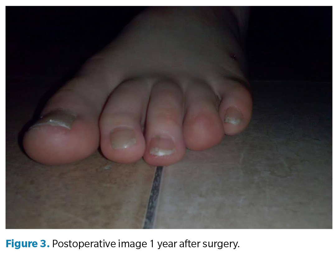
Surgical closure was performed with simple sutures, achieving a satisfactory functional and aesthetic result. A nonadherent dressing and semicompressive bandage were applied. Postoperatively, oral analgesia and weekly follow-up were indicated. Sutures were removed at 14 days, with no complications such as dehiscence, infection, or necrosis. Serial dressings were performed for 4 weeks.
The patient was discharged in September 2024 (12 months after surgery) without signs of recurrence and with good functional and aesthetic adaptation (Figure 3).

Discussion

Osteochondromas are the most common benign bone tumors, representing 20 % to 50 % of all bone tumors. They are superficial bone lesions composed of cortical and medullary bone, covered by hyaline cartilage. Continuity between the cortical and medullary bone of the lesion and the underlying bone is a pathognomonic feature that confirms the diagnosis. Osteochondromas may present as solitary or multiple lesions, the latter being an autosomal dominant condition known as hereditary multiple exostoses. Prevalence has been reported as slightly higher in women (70%), although other studies have not found significant sex differences(5,6,7,8,9).
Most subungual osteochondromas are located in the 1st toe, although cases have been described in the fourth and second toes(2,4,8).
Regarding treatment, Alabdullrahman et al. (2024) proposed that, in most solitary osteochondromas without signs of malignancy, conservative management may be appropriate, highlighting the experimental use of retinoic acid gamma receptor agonists with promising results. In such cases, surgery—whether marginal or wide excision—would be reserved for lesions with suspected malignant transformation(11).
By contrast, Jarolia et al. (2024) advocated surgery as the treatment of choice, noting that resection accompanied by thorough bone curettage is essential to reduce recurrence rates. They also emphasized the importance of submitting the surgical specimen for histopathological analysis to differentiate osteochondromas from other exostoses and to rule out malignancy, particularly in subungual lesions, which may be underdiagnosed(12).
Vázquez-Flores et al. (2004) concurred on the need for surgical resection, using in their series of 27 cases the Dubois-type technique to expose and resect the lesion from its base, followed by meticulous curettage of the bone bed. They also underscored the relevance of precise clinical-radiological diagnosis to differentiate osteochondroma from other subungual masses(13).
Dabrowski et al. (2003), in a study of 74 patients, used a “fish-mouth” incision followed by burr curettage, identifying a higher risk of recurrence in patients younger than 18 years and in those with greater postoperative pain, although no statistically significant association was found with sex, symptom duration, preoperative pain, tumor size, or nail plate destruction(14).
Although the literature often recommends a conservative approach with preservation of the phalanx and exhaustive curettage, complete resection of the distal phalanx was chosen in this case. This decision was based on total bone involvement, recurrent infections, severe nail dystrophy, and radiological and histopathological findings that, although not diagnostic of malignancy, justified a wider surgical margin to minimize recurrence risk.
In conclusion, surgical treatment of osteochondromas is essential due to the potential malignant transformation of these lesions and associated complications, such as compression of adjacent neurovascular structures and functional impairment. Complete surgical resection of the osteochondroma not only eliminates pain and prevents pathologic fractures but also reduces the risk of malignancy, particularly in rapidly growing osteochondromas or those located in atypical areas. Moreover, surgical intervention can significantly improve patients’ quality of life by restoring mobility and normal function of the affected limb.

Conflict of interest
None declared

Funding
None declared

Contributions of the authors
Concept and design: ARM. Data collection: RRR, SSM. Drafting of the manuscript: RRM, RGP. Final review: AMRP

References

  1. Hwang S, Kim M, Park HJ. Subungual osteochondroma. Indian J Dermatol Venereol Leprol. 2017;83(5):620-1. DOI: 10.4103/ijdvl.IJDVL_931_16.
  2. Tamayo-Pacho F, Mora-Ríos FG, Mejía-Rohenes LC, Montero-Quijano MG, López-Marmolejo A. Osteoosteocondromas: presentación del osteoosteocondroma subungueal. Acta Ortop Mex. 2017;31(4):162-4.
  3. Rogozhin DV, Bulycheva IV, Kushlinsky NE, Konovalov DM, Talalaev AG, Roshchin VY, et al. Osteochondroma in children and adolescents. Arkh Patol. 2015;77(3):37-40. DOI: 10.17116/patol201577337-40.
  4. Caino S, Alba R, Bevilacqua S, Roizen M, Obregón MG, Fano V. Osteochondromatosis: clinical variability and factors related to quality of life in children and adults. Arch Argent Pediatr. 2022;120(3):180-6.
  5. Mordenti M, Shih F, Boarini M, Pedrini E, Gnoli M, Antonioli D, et al. The natural history of multiple osteochondroma in a large Italian cohort of pediatric patients. Bone. 2020;139:115499. DOI: 10.1016/j.bone.2020.115499.
  6. Kose O, Ertas A, Celiktas M, Kisin B. Fracture of an osteochondroma treated successfully with total excision: Two case reports. Cases J. 2009;2:8062. D DOI: 10.4076/1757-1626-2-8062.
  7. DaCambra MP, Gupta SK, Ferri-de-Barros F. Subungual exostosis of the toes: A systematic review. Clin Orthop Relat Res. 2014;472(4):1251-9. DOI: 10.1007/s11999-013-3345-4.
  8. Ding W, Han T, Gu J, Xue X. Bizarre parosteal osteochondromatous proliferation of the metatarsal bone: A case report. Asian J Surg. 2024;47(2):1195-6. DOI: 10.1016/j.asjsur.2023.11.023.
  9. Oh S, Won SH, Kim WS, Park MS, Sung KH. Lower extremity deformity and its risk factors in patients with solitary osteochondroma. J Orthop Surg Res. 2024;19(1):415. DOI: 10.1186/s13018-024-04908-4.
  10. García-Ramos CL, Buganza-Tepole M, Obil-Chavarría CA, Reyes-Sánchez AA. Osteoosteocondroma espinal: diagnóstico por imagen y tratamiento. Reporte de casos. Cir Cir. 2015;83(6):496-500.
  11. Alabdullrahman LW, Mabrouk A, Byerly DW. Osteochondroma. En: StatPearls. Treasure Island (FL): StatPearls Publishing; 2025.
  12. Jarolia M, Mlv SK, Digge VK, Panda AK. Subungual osteochondroma of the great toe: a case report. J Am Podiatr Med Assoc. 2024;114(2):22-8. DOI: 10.7547/22-208.
  13. Vázquez-Flores H, Domínguez-Cherit J, Vega-Memije ME, Sáez-De-Ocariz M. Subungual osteochondroma: Clinical and radiologic features and treatment. Dermatol Surg. 2004;30(7):1031-4. DOI: 10.1097/00042728-200407000-00013.
  14. Dabrowski M, Rusek D, Da?czak-Pazdrowska A, Litowi?ska A. The influence of clinical factors on treatment outcome and a recurrence of surgically removed protruded subungual osteochondroma and subungual exostosis. J Clin Med. 2023;12(19):6413. DOI: 10.3390/jcm12196413.

Nuevo comentario

Security code:
CAPTCHA code image
Speak the codeChange the code
 

Comentarios

No comments in this article

Bibliografía

1. Hwang S, Kim M, Park HJ. Subungual osteochondroma. Indian J Dermatol Venereol Leprol. 2017;83(5):620-1.
2. Tamayo-Pacho F, Mora-Ríos FG, Mejía-Rohenes LC, Montero-Quijano MG, López-Marmolejo A. Osteoosteocondromas: presentación del osteoosteocondroma subungueal. Acta Ortop Mex. 2017;31(4):162-4.
3. Rogozhin DV, Bulycheva IV, Kushlinsky NE, Konovalov DM, Talalaev AG, Roshchin VY, et al. [Osteochondroma in children and adolescents]. Arkh Patol. 2015;77(3):37-40.
4. Caino S, Alba R, Bevilacqua S, Roizen M, Obregón MG, Fano V. Osteochondromatosis: clinical variability and factors related to quality of life in children and adults. Arch Argent Pediatr. 2022;120(3):180-6.
5. Mordenti M, Shih F, Boarini M, Pedrini E, Gnoli M, Antonioli D, et al. The natural history of multiple osteochondroma in a large Italian cohort of pediatric patients. Bone. 2020;139:115499.
6. Kose O, Ertas A, Celiktas M, Kisin B. Fracture of an osteochondroma treated successfully with total excision: Two case reports. Cases J. 2009;2:8062. D
7. DaCambra MP, Gupta SK, Ferri-de-Barros F. Subungual exostosis of the toes: A systematic review. Clin Orthop Relat Res. 2014;472(4):1251-9.
8. Ding W, Han T, Gu J, Xue X. Bizarre parosteal osteochondromatous proliferation of the metatarsal bone: A case report. Asian J Surg. 2024;47(2):1195-6.
9. Oh S, Won SH, Kim WS, Park MS, Sung KH. Lower extremity deformity and its risk factors in patients with solitary osteochondroma. J Orthop Surg Res. 2024;19(1):415.
10. García-Ramos CL, Buganza-Tepole M, Obil-Chavarría CA, Reyes-Sánchez AA. Osteoosteocondroma espinal: diagnóstico por imagen y tratamiento. Reporte de casos. Cir Cir. 2015;83(6):496-500.
11. Alabdullrahman LW, Mabrouk A, Byerly DW. Osteochondroma. En: StatPearls. Treasure Island (FL): StatPearls Publishing; 2025.
12. Jarolia M, Mlv SK, Digge VK, Panda AK. Subungual osteochondroma of the great toe: a case report. J Am Podiatr Med Assoc. 2024;114(2):22-8.
13. Vázquez-Flores H, Domínguez-Cherit J, Vega-Memije ME, Sáez-De-Ocariz M. Subungual osteochondroma: Clinical and radiologic features and treatment. Dermatol Surg. 2004;30(7):1031-4.
14. Dąbrowski M, Rusek D, Dańczak-Pazdrowska A, Litowińska A. The influence of clinical factors on treatment outcome and a recurrence of surgically removed protruded subungual osteochondroma and subungual exostosis. J Clin Med. 2023;12(19):6413.

Artículos relacionados

Clinical and functional effectiveness of posterior leg muscle lengthening: a systematic review

Rev. Esp. Pod. 2025; 36(2): 176-183 / DOI: 10.20986/revesppod.2025.1747/2025

Thermography for diagnosis and monitoring of peripheral artery disease: a narrative review

Rev. Esp. Pod. 2025; 36(2): 170-175 / DOI: 10.20986/revesppod.2025.1741/2025

Consequences of juvenile dermatomyositis at the podiatric level: a literature review

Rev. Esp. Pod. 2025; 36(2): 164-169 / DOI: 10.20986/revesppod.2025.1731/2025

Podiatric implications of Guillain-Barré syndrome sequelae: a systematic review

Rev. Esp. Pod. 2025; 36(2): 156-163 / DOI: 10.20986/revesppod.2025.1742/2025

Systematic review of podiatric involvement in Andrade syndrome

Rev. Esp. Pod. 2025; 36(2): 148-155 / DOI: 10.20986/revesppod.2025.1740/2025

Surgical management of intratendinous multinodular ganglion. A clinical case

Rev. Esp. Pod. 2025; 36(2): 143-147 / DOI: 10.20986/revesppod.2025.1755/2025

Surgical treatment of first toe necrosis due to septicemia: clinical case

Rev. Esp. Pod. 2025; 36(2): 135-138 / DOI: 10.20986/revesppod.2025.1746/2025

Relationship between gait parameters and fear of falling in individuals with fibromyalgia

Rev. Esp. Pod. 2025; 36(2): 103-110 / DOI: 10.20986/revesppod.2025.1745/2025

Influence of climbing-shoe size on the podiatric conditions of the climber’s foot: an observational study

Rev. Esp. Pod. 2025; 36(2): 91-95 / DOI: 10.20986/revesppod.2025.1733/2025

Effectiveness of the use of bone cement in the treatment of diabetic foot osteomyelitis

Rev. Esp. Pod. 2025; 36(1): 67-74 / DOI: 10.20986/revesppod.2025.1718/2024

Molluscum pendulum on the foot. Clinical case

Rev. Esp. Pod. 2025; 36(1): 54-58 / DOI: 10.20986/revesppod.2025.1728/2025

Epidermoid carcinoma associated to onichocryptosis: a case report

Rev. Esp. Pod. 2025; 36(1): 48-53 / DOI: 10.20986/revesppod.2025.1729/2025

Determining variables in foot pain in padel players. Observational analysis

Rev. Esp. Pod. 2025; 36(1): 35-41 / DOI: 10.20986/revesppod.2025.1727/2025

Treatment of plantar fascitis with intratissue percutaneous electrolysis. Systematic review and metaanalisis

Rev. Esp. Pod. 2025; 36(1): 9-16 / DOI: 10.20986/revesppod.2025.1711/2024

Comparative study of three techniques for the ring block of the hallux: Frost´s H and modifications

Rev. Esp. Pod. 2025; 36(1): 3-8 / DOI: 10.20986/revesppod.2025.1709/2024

Brachydactyly type B: rare case report

Rev. Esp. Pod. 2024; 35(2): 116-119 / DOI: 10.20986/revesppod.2024.1702/2024

Ultrasound-guided percutaneous needle fasciotomy. Case series

Rev. Esp. Pod. 2024; 35(2): 105-109 / DOI: 10.20986/revesppod.2024.1710/2024

Injuries in the foot and ankle region in rugby players from the University of Malaga. Observational study

Rev. Esp. Pod. 2024; 35(2): 85-90 / DOI: 10.20986/revesppod.2024.1700/2024

Anatomy and sonopathology of the metatarsophalangeal plantar plate

Rev. Esp. Pod. 2024; 35(1): 57-68 / DOI: 10.20986/revesppod.2024.1693/2024

Eficacy of arthroscopy in hallux rigidus: a sistematic review

Rev. Esp. Pod. 2024; 35(1): 49-56 / DOI: 10.20986/revesppod.2024.1689/2024

Qualitative pilot study on the orthotic management of tibialis posterior tendon dysfunction by podiatrists

Rev. Esp. Pod. 2024; 35(1): 30-35 / DOI: 10.20986/revesppod.2024.1686/2024

Surgical approach to rheumatic foot. Case series

Rev. Esp. Pod. 2024; 35(1): 17-21 / DOI: 10.20986/revesppod.2024.1678/2023

Ultrasound-detected ankle tenosynovitis is associated with foot function disorders: a cross-sectional study

Rev. Esp. Pod. 2024; 35(1): 11-16 / DOI: 10.20986/revesppod.2024.1692/2024

Measuring tools for nail pathology: literature review

Rev. Esp. Pod. 2023; 34(2): 113-120 / DOI: 10.20986/revesppod.2023.1667/2023

Podiatrically significant injuries in the regular practice of contact sports.

Rev. Esp. Pod. 2023; 34(2): 104-108 / DOI: 10.20986/revesppod.2023.1668/2023

Rotational anomalies in women with hallux valgus. Observational study

Rev. Esp. Pod. 2023; 34(2): 90-98 / DOI: 10.20986/revesppod.2023.1673/2023

Morphology and function of the foot in patients with sciatica. A cross-sectional study

Rev. Esp. Pod. 2023; 34(2): 82-89 / DOI: 10.20986/revesppod.2023.1672/2023

Limitation of ankle dorsiflexion and hallux in master category swimmers

Rev. Esp. Pod. 2023; 34(2): 74-81 / DOI: 10.20986/revesppod.2023.1671/2023

Affections of the lower limb in subjects that perform kick-boxing: descriptive study

Rev. Esp. Pod. 2023; 34(2): 69-73 / DOI: 10.20986/revesppod.2023.1669/2023

Treatment for plantar fasciitis with biomechanical socks. Preliminary results of a randomized clinical trial

Rev. Esp. Pod. 2023; 34(2): 62-68 / DOI: 10.20986/revesppod.2023.1663/2023

Sonographic mapping of the nerves of the medial side of the ankle and foot

Rev. Esp. Pod. 2023; 34(1): 52-57 / DOI: 10.20986/revesppod.2023.1665/2023

Surgical treatment of diabetic foot plantar ulcer. Report of a clinical case

Rev. Esp. Pod. 2023; 34(1): 47-51 / DOI: 10.20986/revesppod.2023.1658/2023

Thermography of the vasodilator effect of local anaesthetics in ingrown toenail surgery

Rev. Esp. Pod. 2023; 34(1): 32-34 / DOI: 10.20986/revesppod.2023.1660/2023

Influence of surgical treatment of hallux abducto-valgus on first ray mobility

Rev. Esp. Pod. 2023; 34(1): 13-18 / DOI: 10.20986/revesppod.2023.1650/2022

Estimation of the ossification of the bones of the foot about the population of Extremadura. Observational study

Rev. Esp. Pod. 2023; 34(1): 3-12 / DOI: 10.20986/revesppod.2023.1654/2022

Surgical resolution of an hallucal interphalangeal sesamoid

Rev. Esp. Pod. 2022; 33(2): 122-127 / DOI: 10.20986/revesppod.2022.1645/2022

Reduction of dynamic plantar pressures in the medial plantar forefoot with biomechanical socks

Rev. Esp. Pod. 2022; 33(2): 110-114 / DOI: 10.20986/revesppod.2022.1646/2022

A new modification of the Winograd technique for the treatment of ingrown toenail

Rev. Esp. Pod. 2022; 33(2): 104-109 / DOI: 10.20986/revesppod.2022.1634/2022

Effect of custom rigid insoles in compressive plantar heel pain: a prospective case series

Rev. Esp. Pod. 2022; 33(2): 96-103 / DOI: 10.20986/revesppod.2022.1647/2022

Plantar pressues in two sock models with different three-dimensional wave separation

Rev. Esp. Pod. 2022; 33(2): 88-95 / DOI: 10.20986/revesppod.2022.1641/2022

Application of hyaluronic acid in the treatment of foot ulcers

Rev. Esp. Pod. 2022; 33(2): 77-78 / DOI: 10.20986/revesppod.2022.1648/2022

Update of new neuromuscular bandage techniques in podiatry

Rev. Esp. Pod. 2022; 33(1): 59-65 / DOI: 10.20986/revesppod.2022.1630/2022

False rheumatoid nodule in the left third toe and its surgical treatment. A case report

Rev. Esp. Pod. 2022; 33(1): 54-58 / DOI: 10.20986/revesppod.2022.1628/2021

Clinical case: angioleiomyoma on the foot

Rev. Esp. Pod. 2022; 33(1): 50-53 / DOI: 10.20986/revesppod.2022.1622/2021

Pain and foot posture in patients with systemic lupus erythematosus. A descriptive study

Rev. Esp. Pod. 2022; 33(1): 35-40 / DOI: 10.20986/revesppod.2022.1633/2022

Baropodometric variation with weight loss: an experimental study

Rev. Esp. Pod. 2022; 33(1): 21-28 / DOI: 10.20986/revesppod.2022.1627/2021

Effectiveness of collagen injection for plantar fasciitis: randomised clinical trial

Rev. Esp. Pod. 2022; 33(1): 14-20 / DOI: 10.20986/revesppod.2022.1608/2021

Comparative study between two techniques of local troncular anesthesia of the first toe

Rev. Esp. Pod. 2022; 33(1): 9-13 / DOI: 10.20986/revesppod.2022.1605/2021

Timing of foot stance in relation to the child’s age: a descriptive observational study

Rev. Esp. Pod. 2022; 33(1): 3-8 / DOI: 10.20986/revesppod.2022.1589/2020

Longitudinal arch load-sharing system of the foot

Rev Esp Podol. 2017;28(1):37-45

Talectomy. A case report

Rev. Esp. Pod. 2021; 32(2): 140-145 / DOI: 10.20986/revesppod.2021.1616/2021

Biomechanical study test in children and adolescents: a systematic review

Rev. Esp. Pod. 2021; 32(2): 132-139 / DOI: 10.20986/revesppod.2021.1611/2021

Narrative review of the use and applications of radiofrequency for the treatment of musculoskeletal pain

Rev. Esp. Pod. 2021; 32(2): 123-131 / DOI: 10.20986/revesppod.2021.1601/2021

Symmetry in podal temperature in diabetic patients and its relationship with the postural index of the foot

Rev. Esp. Pod. 2021; 32(2): 106-115 / DOI: 10.20986/revesppod.2021.1607/2021

Kinematic analysis of ankle and midfoot joints in children with osteogenesis imperfecta

Rev. Esp. Pod. 2021; 32(2): 99-105 / DOI: 10.20986/revesppod.2021.1603/2021

Reduction of dynamic plantar pressures in an experimental sock. A preliminary report

Rev. Esp. Pod. 2021; 32(2): 86-92 / DOI: 10.20986/revesppod.2021.1619/2021

Foot temperature in the different phases of the menstrual cycle

Rev. Esp. Pod. 2021; 32(2): 79-85 / DOI: 10.20986/revesppod.2021.1606/2021

Effect of laser therapy on post surgical pain and edema after hallux valgus surgery: a prospective analytical study

Rev. Esp. Pod. 2021; 32(2): 74-78 / DOI: 10.20986/revesppod.2021.1570/2020

Morton’s neuroma decompression: current insights & recommendations

Rev. Esp. Pod. 2021; 32(1): 63-69 / DOI: 10.20986/revesppod.2021.1609/2021

Multibranched acquired digital fibrokeratoma. Clinical case

Rev. Esp. Pod. 2021; 32(1): 58-62 / DOI: 10.20986/revesppod.2021.1578/2020

Relationship of the design of football boots to the injury in football: systematic review

Rev. Esp. Pod. 2021; 32(1): 49-57 / DOI: 10.20986/revesppod.2021.1580/2020

Effectiveness of conservative treatments in patients with calcaneal apophysitis. Systematic review

Rev. Esp. Pod. 2021; 32(1): 42-48 / DOI: 10.20986/revesppod.2021.1593/2020

Radiographic study of the mobility of the first ray in the sagittal and frontal planes

Rev. Esp. Pod. 2021; 32(1): 27-35 / DOI: 10.20986/revesppod.2021.1600/2021

Descriptive analysis of 3D foot motion using inertial sensors: comparison between lower extremities

Rev. Esp. Pod. 2021; 32(1): 13-17 / DOI: 10.20986/revesppod.2021.1594/2020

Relationship between dorsal ankle flexion and medial column flexibility

Rev. Esp. Pod. 2021; 32(1): 7-12 / DOI: 10.20986/revesppod.2021.1572/2020

Meschan angle through clinical photography: pilot study

Rev. Esp. Pod. 2021; 32(1): 2-6 / DOI: 10.20986/revesppod.2021.1565/2020

Surgical reconstruction of a complex neuroarthopatic charcot foot: a case report

Rev. Esp. Pod. 2020; 31(2): 117-125 / DOI: 10.20986/revesppod.2020.1569/2020

Quality of life in patients with hallux valgux assessed by SF-36, SF-12 and FHSQ questionnaires: a systematic review

Rev. Esp. Pod. 2020; 31(2): 102-109 / DOI: 10.20986/revesppod.2020.1583/2020

Distal metatarsal mini-invasive osteotomy for the iatrogenic metatarsalgia. Retrospective study of a serie of cases

Rev. Esp. Pod. 2020; 31(2): 93-101 / DOI: 10.20986/revesppod.2020.1574/2020

Anxiety study in the presurgical patient in foot surgery

Rev. Esp. Pod. 2020; 31(2): 87-92 / DOI: 10.20986/revesppod.2020.1562/2020

General and podiatric quality of life in postmenopausal women with bone metabolism disorders: observational study

Rev. Esp. Pod. 2020; 31(2): 76-81 / DOI: 10.20986/revesppod.2020.1559/2020

Study of the response to aerobic resistance exercises based on the type of foot in the child population

Rev. Esp. Pod. 2020; 31(2): 71-75 / DOI: 10.20986/revesppod.2020.1560/2020

Dynamic instability of the pelvis and its relation to plantar pressures in runners

Rev. Esp. Pod. 2020; 31(2): 65-70 / DOI: 10.20986/revesppod.2020.1558/2020

The impact of the foot for the health

Rev. Esp. Pod. 2020; 31(2): 63-64 / DOI: 10.20986/revesppod.2020.1586/2020

Multidisciplinary approach to neuroischaemic ulcers; a case report

Rev. Esp. Pod. 2020; 31(1): 55-61 / DOI: 10.20986/revesppod.2020.1573/2020

How to give advise on footwear to running patients? From scientific evidence to clinical experience

Rev. Esp. Pod. 2020; 31(1): 46-54 / DOI: 10.20986/revesppod.2020.1571/2020

Effects of a stabilizer sock in balance in patients with different neurologic diseases

Rev. Esp. Pod. 2020; 31(1): 38-45 / DOI: 10.20986/revesppod.2020.1566/2020

Analysis of stiffness of the first ray during weightbearing with a new device: a feasibility exploratory study

Rev. Esp. Pod. 2020; 31(1): 24-30 / DOI: 10.20986/revesppod.2020.1555/2019

Study of the effectiveness of foot orthoses in frequent foot pathologies

Rev. Esp. Pod. 2020; 31(1): 16-23 / DOI: 10.20986/revesppod.2020.1557/2020

Minimally invasive surgery of the lesser toes

Rev. Esp. Pod. 2019; 30(2): 113-126 / DOI: 10.20986/revesppod.2019.1546/2019

Ecoguided puncture in front of the blind puncture in the nervous lock of the foot: narrative bibliographical review

Rev. Esp. Pod. 2019; 30(2): 101-108 / DOI: 10.20986/revesppod.2019.1548/2019

Provision of orthopaedic footwear to patients in a hospital area: transversal study

Rev. Esp. Pod. 2019; 30(2): 81-86 / DOI: 10.20986/revesppod.2019.1547/2019

Effects of the Low-Dye tape on the foot. A narrative review

Rev. Esp. Pod. 2019; 30(1): 38-52 / DOI: 10.20986/revesppod.2019.1526/2018

Extraction of gotous tofos. Clinical case.

Rev. Esp. Pod. 2019; 30(1): 24-30 / DOI: 10.20986/revesppod.2019.1515/2018

Normative values to determine a flatfoot or cavus foot

Rev. Esp. Pod. 2019; 30(1): 15-23 / DOI: 10.20986/revesppod.2019.1540/2019

Tissue stress model. Clinical applications in foot pathology

Rev. Esp. Pod. 2018; 29(2): 101-112 / DOI: 10.20986/revesppod.2018.1531/2018

S-Plasty for excision of a circular lession of the hallux: a case report

Rev. Esp. Pod. 2018; 29(2): 94-100 / DOI: 10.20986/revesppod.2018.1525/2018

Osteochondroma or subungual exostosis subungueal? A systematic review

Rev. Esp. Pod. 2018; 29(2): 81-86 / DOI: 10.20986/revesppod.2018.1521/2018

Frequency of recurring onychocryptosis in diabetic population derived to a podiatric clinic

Rev. Esp. Pod. 2018; 29(2): 75-80 / DOI: 10.20986/revesppod.2018.1523/2018

Flatfoot surgery: a personal view

Rev Esp Podol. 2018;29(1):49-59 / DOI: 10.20986/revesppod.2018.1516/2018

Effectiveness of conservative orthotic treatment in flexible pediatric flatfoot

Rev Esp Podol. 2018;29(1):2-12 / DOI: 10.20986/revesppod.2018.1507/2018

Instrucciones para citar

Rayo Martín A, Rayo Martín R, Rayo Rosado R, Sánchez-Morilla S, García de la Peña R, Rayo Pérez A, et all. Ostechondroma in the distal phalanx of a pediatric patient: a case report. Rev Esp Pod 2025; 36(2): 139-142 / DOI: 1020986/revesppod20251751/2025


Descargar a un gestores de citas

Descargue la cita de este artículo haciendo clic en uno de los siguientes gestores de citas:

Métrica

Este artículo ha sido visitado 23 veces.
Este artículo ha sido descargado 0 veces.

Estadísticas de Dimensions


Estadísticas de Plum Analytics

Ficha Técnica

Recibido: 23/07/2025

Aceptado: 25/09/2025

Prepublicado: 24/11/2025

Publicado: 31/01/2026

Tiempo de revisión del artículo: 63 días

Tiempo de prepublicación: 124 días

Tiempo de edición del artículo: 192 días


Compartir

Este artículo aun no tiene valoraciones .
Reader rating:
Rate this article:
2026 Revista Española de Podología
ISSN: 0210-1238   e-ISSN: 2695-463X

      Indexada en: