DOI: http://dx.doi.org/10.20986/revesppod.2024.1692/2024
ORIGINAL
La tenosinovitis ecográfica de tobillo está asociada a la alteración funcional del pie: un estudio transversal
Ultrasound-detected ankle tenosynovitis is associated with foot function disorders: a cross-sectional study
Anna Tribó Crespo1
César Díaz Torné2
Patricia Moya Alvarado2
Francesc Monés Serrano3
1Dpto. de Biomecànica. Centre Podològic Monés. Badalona, Barcelona, España.
2Servei de Reumatologia. Hospital de la Santa Creu y Sant Pau. Barcelona, Barcelona.
3Servei d’Endocrinologia i Nutrició. Hospital Germans Trias i Pujol. Badalona, Barcelona, España
Resumen
Introducción: Este estudio tiene como objetivo explorar la asociación de los trastornos estáticos y dinámicos del pie con la tendinopatía ecográfica del tobillo, más específicamente del tendón del tibial posterior y los tendones peroneos del tobillo, en sujetos sanos.
Pacientes y métodos: Se diseñó un estudio descriptivo, analítico, transversal que se llevó a cabo en la Unidad de Reumatología del Hospital de la Santa Creu i Sant Pau, Barcelona. Se registró la edad, sexo, índice de postura del pie (FPI-6), índice cinético del pie (FKI) y la característica patológica ecográfica, del tendón del tibial posterior y de los tendones peroneos. Se utilizó la regresión logística para evaluar la probabilidad de que los trastornos posturales y/o funcionales del pie estén relacionados con los hallazgos ecográficos de patología.
Resultados: La asociación entre las características ecográficas de tenosinovitis y el trastorno dinámico resultó estadísticamente significativa (p = 0.00). La asociación entre la afección ecográfica y los trastornos simultáneos, estáticos y dinámicos, del pie también fue estadísticamente significativa (p = 0.03). La hiperpronación dinámica se asocia a la tenosinovitis del tendón tibial posterior (p = 0.012). Se realizó un análisis complementario, de carácter exploratorio, para valorar la probabilidad de asociación entre trastorno dinámico y tendinopatía de tobillo que mostró un odds ratio de 5.9.
Conclusiones: Este estudio transversal informa sobre la asociación entre trastornos de la función del pie y características ecográficas de tobillo. El FKI destaca como diagnóstico numérico capaz de detectar factores de riesgo de patología inflamatoria del tobillo.
Palabras clave: Análisis cinético, presión plantar, estudio de la marcha, tendinopatía, tenosinovitis de tobillo, ecografía de pie
Abstract
Introduction: This study aims to explore the association of static and dynamic foot disorders with ankle tendinopathy measured by ultrasound, more specifically of tibialis posterior tendon and peroneal ankle tendons, in healthy subjects.
Patients and methods: A cross-sectional descriptive and analytical study was designed and carried out in Rheumatology Unit, Hospital de la Santa Creu i Sant Pau, Barcelona. For each participant were registered: age, sex, Foot Posture Index (FPI-6), Foot Kinetic Index (FKI) and tibialis posterior and peroneals tendons ultrasound patological features. Logistic regression was used to evaluate the likelihood that postural or/and functional foot disorders is related to ankle sonographic features.
Results: Association between ultrasound tenosynovitis features and dynamic disorder resulted statistically significant (p = 0.00). Association between ultrasound affection and simultaneous, static and dynamic, foot disorders was also statistically significant (p = 0.03). Dynamic’s overpronation is associated to tibialis posterior tendon tenosynovitis (p = 0.012). Dynamic disorders were associated with the likelihood of ankle tendinopathy in unadjusted models (OR = 5.9).
Conclusions: This cross-sectional study reports association between foot function disorders and ankle sonographic features. It FKI highlights as a diagnostic score able to detect risk factors for inflammatory ankle pathology.
Keywords: Kinetic assessment, plantar pressures, gait analysis, tendinopathy, ankle tenosynovitis, foot ultrasound
Correspondence: Anna Tribó Crespo
annatribo00@gmail.com
Recibido: 11/03/2024
Aceptado: 10/04/2024
Introducción
La teoría del estrés tisular explica que antes de una lesión o fractura de cualquier tejido, orgánico o no, sometido a cierto esfuerzo, este experimentará una serie de cambios reversibles dentro del área de deformidad elástica, o irreversibles si ya se encuentra dentro de su área de deformidad plástica, aun eliminando el estrés o sufrimiento(1). Estos cambios, que se producen en el interior de los tejidos anatómicos, como músculos o tendones(2), son progresivos: hipertrofia, inflamación (con aumento del fluido intersticial, visualizado específicamente en los tendones del interior de las vainas), heterogeneidad fibrilar y finalmente rotura parcial o completa. Todos estos cambios pueden verse en la ecografía, lo cual requiere una amplia curva de aprendizaje para el evaluador, si bien es un método de diagnóstico válido. Parece lógico que, para prevenir lesiones, debamos identificar primero el esfuerzo o estrés de una estructura determinada para poder evitarlo(3).
Específicamente, si evaluamos patologías de los músculos y tendones estabilizadores del tobillo, debemos poder usar sistemas para detectar su sufrimiento por sobrestimulación. La exploración visual de la postura del paciente proporciona información para la evaluación estática, sin movimiento, pero no durante la marcha, ya que los movimientos ejecutados se realizan a una frecuencia mayor de la que el ojo humano es capaz de detectar. Los sistemas de evaluación biomecánica de tobillo y pie pueden ser cinemáticos(4) (con cámaras de alta frecuencia con extracción de datos en 3D) o cinéticos (basados en registros de fuerza o presión utilizando elementos como plataformas dinamométricas o sensores de presión). Para la valoración estática del sujeto, solo se ha validado el índice de postura del pie (FPI-6). Para la valoración dinámica se han empleado diferentes variables, tanto en la exploración cinética como cinemática, si bien no se ha llegado a un consenso o validez de ningún método o valor de diagnóstico. Se enfatiza que el coste económico requerido por un laboratorio de escaneo cinemático (cámaras externas de alta frecuencia y distribución para reconstrucción 3D) es mayor que el que se necesita para la realización de escáneres cinéticos (plataformas de presión o plantillas de sensores).
Los trastornos biomecánicos, tanto estáticos como dinámicos, parecen ser los responsables del estrés de los tejidos en los músculos estabilizadores del tobillo(5). Nuestro objetivo fue conocer con qué frecuencia eso es cuantitativamente real. En este sentido, nuestro resultado fue valorar la asociación entre la marcha patológica y el estrés tisular en el tobillo. Fue necesario elegir un valor de valoración postural y una variable de valoración dinámica, ambos capaces de detectar trastornos, con el objetivo de relacionarlos con la posible enfermedad inflamatoria.
Es necesario conocer la importancia de ciertos factores con relación a la aparición, o no, de una determinada patología para decidir si estudiarlos, o no, en futuros proyectos como posibles factores de riesgo. El hecho de no haber encontrado referencias bibliográficas sobre la relación entre estas tres observaciones en particular (trastorno postural, trastorno dinámico y afectación tendinosa), nos llevó a diseñar un estudio observacional descriptivo y analítico, con naturaleza exploratoria. El resultado general del estudio fue explorar la asociación que existe entre los trastornos dinámicos y estáticos del pie y la tendinopatía del tobillo, específicamente del tendón tibial posterior y de los tendones peroneos. Como objetivos específicos, intentamos describir la frecuencia de la variable de trastorno postural para sus tres posibles categorías (pronado, supinado y neutro), así como la frecuencia de la variable de trastorno dinámico, para sus tres posibles categorías (pronado, supinado y neutro), y también describir la frecuencia de las variables de enfermedad inflamatoria, mediante ecografías del tendón del tibial posterior y tendones peroneos. Luego intentamos determinar la relación estadística, fuerza y riesgo relativo entre las tres posibles variables y sus diferentes posibles categorías.
Pacientes y métodos
Este es un estudio descriptivo y analítico transversal. Los datos se recopilaron entre enero y agosto de 2017, en la ciudad de Barcelona. Los participantes en el estudio fueron reclutados entre estudiantes, profesionales, socios y familiares cercanos al “Hospital de Sant Pau i la Santa Creu” de Barcelona. Los participantes fueron reclutados de acuerdo con los siguientes criterios de selección: ser mayor de 18 años, no tener síntomas clínicos, ausencia de antecedentes de patologías osteoarticulares, antecedentes traumáticos o quirúrgicos de pie y tobillo. Se excluyó a aquellos participantes que utilizaron tratamientos con plantillas para los pies durante el último año(6).
Se analizaron 55 participantes. El tamaño total de la muestra fue de 104 pies válidos. Cada pie fue valorado de forma independiente, aunque perteneciera al mismo participante(7).
El tamaño de la muestra se determinó de manera no probabilística, por conveniencia para fines exploratorios. Se destaca que el tamaño de la muestra tiene suficiente poder estadístico para responder a los objetivos.
Este estudio se llevó a cabo de acuerdo con las recomendaciones contenidas en la “Declaración para mejorar la presentación de estudios observacionales en epidemiología”, STROBE, Fortalecimiento de la Presentación de Estudios Observacionales en Epidemiología.
Herramientas y procedimientos
Todo hombre o mujer mayor de 18 años, sin síntomas en las extremidades inferiores, que tuviera alguna relación con el Departamento de Reumatología, fue candidato para el estudio. En una única visita para cada participante, se recopilaron los siguientes datos: características demográficas, edad, peso, altura y tratamientos médicos o podológicos, valoración postural y dinámica y ecografías del tobillo.
Valoración postural
Para valorar los trastornos posturales del pie, cada participante fue examinado en posición estática y se determinó el valor del índice de postura del pie-6 (FPI-6) )(8,9)para cada pie. Esta entrevista y la primera evaluación siempre fueron realizadas por el mismo examinador, un podólogo con más de dos años de experiencia clínica que procedió a rellenar una hoja de recopilación de datos. Esta recogida de datos fue ciega de los datos clínicos, del estudio dinámico (FKI) y de las ecografías del tobillo.
El FPI-6 es un índice validado de evaluación estática de la postura del pie para la determinación cuantitativa de la postura del paciente, mediante la observación y consecuente puntuación de 6 criterios. El FPI-6 tiene en cuenta el antepié, retropié y mediopié en los tres planos anatómicos. El sistema de puntuación utilizado es de 5 puntos de la escala tipo Likert (de +2 a -2), donde la puntuación positiva máxima se otorgará a los criterios de mayor pronación y los valores negativos máximos a los de mayor supinación. En este sentido, el FPI se obtiene sumando las puntuaciones (-2, -1, 0, 1 o 2) dadas a cada uno de los seis criterios. Según los resultados, se clasifica como pie neutral (de 0 a 5), pronado (de 6 a 9), altamente pronado (de 10 a 12), supinado (de -1 a -4) y altamente supinado (de -5 a -12).
Valoración dinámica
Para la evaluación de trastornos dinámicos, se utilizó el índice cinético del pie (FKI). Para ello, cada participante caminaba descalzo a una velocidad cómoda. El sujeto podía caminar libremente en línea recta, en la que el grosor de la plataforma no interfiriera, en ambas direcciones, durante aproximadamente 6 metros, y realizar un mínimo de 5 repeticiones, en una habitación cómoda y ligeramente iluminada. Las presiones dinámicas del participante durante el análisis de la marcha fueron registradas utilizando una alfombra de sensores capacitivos de 49 x 49 cm (AmCube, Gargas, Francia). Se evaluaron la trayectoria del Centro de Presiones (Cop)(10,11,12) y la línea de puntos de presión máxima utilizando el software FootWorkPro, de AMCube. Se registraron varios pasos para cada pie, pero solo se guardaron los tres pasos más representativos. A partir de estos, se obtuvo el valor del índice cinético del pie (FKI). El proceso de registro y puntuación de los diferentes ítems para obtener el FKI siempre fue realizado por el mismo evaluador, un podólogo con más de quince años de experiencia clínica, y se realizó a ciegas respecto a los datos clínicos, al estudio de la postura (FPI-6) y a la evaluación por ultrasonido.
El índice cinético del pie (FKI) es un índice cuantitativo que se usa para valorar la función dinámica del pie, en base a la comparativa de la trayectoria del centro de presiones (Cop) con la ruta de presión máxima. El FKI es un método de examen clínico, no validado, para la cuantificación de la funcionalidad dinámica del pie a través de la observación y consiguiente puntuación del retropié, mediopié y antepié con respecto a las cuatro fases de la marcha: respuesta a la carga, mitad del apoyo, apoyo terminal y pre-balanceo o fase previa a la oscilación. Se trata un índice que permite puntuar los momentos de fuerza en el plano frontal, pronador y supinador, teniendo también en cuenta su duración. El sistema de puntuación del FKI da una puntuación positiva máxima a los criterios de mayor pronación y unos valores negativos máximos a los de mayor supinación. Según los resultados, se clasifica como pie neutro (de -3 a 3), pronado (de 4 a 12) y supinado (de -4 a -12).
Valoración de la tendinopatía
En cuanto a la valoración por ecografía del tobillo, se utilizó un ecógrafo de General Electric Logic 5 Pro (GE Healthcare, California, Estados Unidos) con un transductor lineal compacto de frecuencia de 10 MHz(13). Para la evaluación de la tendinopatía, se observó la ecoestructura del tendón, clasificándola como homogénea o heterogénea. También se valoró si había o no rotura y, en caso afirmativo, se clasificó como parcial o total. Finalmente, se describió si se observaba un aumento de líquido en las vainas tendinosas, considerando como normal el hallazgo de hasta 1.5 mm de líquido en las vainas, y la identificación de una anchura de acumulación de líquido en las vainas > 1.5 mm como patológica y representativa de inflamación. La presencia de cualquiera de las alteraciones mencionadas se consideró positiva para la variable “tenosinovitis detectada por ecografía”. La ecografía del tobillo se realizó en todos los planos anatómicos. Se tuvieron en cuenta el tendón tibial posterior(14) y los tendones del peroneo largo y corto de los compartimentos medial y lateral del tobillo, respectivamente. Tanto la exploración, la evaluación como el registro de datos fueron realizados por un reumatólogo ecografista con más de ocho años de experiencia, y se realizó a ciegas respecto a los datos clínicos, al estudio de la postura (FPI-6) y al conjunto de datos cinéticos (FKI).
Variables de resultado
Las variables de resultado primarias fueron categóricas: trastorno postural existente o no, trastorno dinámico existente o no, y enfermedad inflamatoria de los tendones tibial posterior y/o peroneos existente o no.
Las variables de resultado secundarias fueron: la edad del paciente, como variable continua, y la clasificación del pie, tanto estática como dinámica, en pie pronado, supinador o neutro, resultando así en tres categorías.
Análisis estadístico
Se utilizaron la base de datos de Excel y el paquete de software estadístico SPSS, 24.0® (IBM®, Estados Unidos). Se realizó un análisis descriptivo de las variables, frecuencias para las categóricas y moda, mediana y desviación estándar para las variables continuas. Para analizar la asociación estadística entre la variable de patología detectada en la ecográfica y las variables de trastornos estáticos o dinámicos, se utilizó la prueba de X2 para tablas de contingencia y el nivel de significación adoptado fue p > 0.05.
Se empleó la regresión logística para calcular la odds ratio (OR) y el intervalo de confianza del 95 % (IC) para la tendinopatía basada en trastornos posturales y/o dinámicos del pie. Se calculó, con carácter exploratorio, para encontrar la fuerza de la asociación estadística y el riesgo relativo entre las observaciones: alteraciones estáticas o dinámicas y patología detectada en la ecografía.
Se empleó la regresión logística binaria no ajustada para calcular la odds ratio entre los trastornos estáticos o dinámicos o ambos (como exposición) y la patología detectada en la ecografía (como resultado). A partir de ahí, se obtuvo la odds ratio (OR) como el riesgo de aparición o no de tendinopatía del tibial posterior y/o peroneal en función de si el participante presentaba o no trastornos estáticos o dinámicos. Para cada modelo de regresión logística binaria, se tuvo en cuenta su bondad de ajuste y los intervalos de confianza, y se verificó la calibración utilizando la prueba de Hosmer-Lemeshow.
Resultados
Se registraron 104 pies de 52 participantes. Las mujeres representaron el 67,3 % de la muestra. La media de edad fue de 42.17 años (19-88).
La frecuencia de aparición de patología detectada en la ecografía entre los 104 pies estudiados fue del 45.2 % (47 pies). Treinta y cinco pies presentaron tenosinovitis del tendón tibial posterior detectada mediante ecografía, 12 pies afectación de los tendones peroneos y 10 pies patología simultánea de ambas estructuras.
La frecuencia de trastorno postural (FPI-6) dentro de la muestra fue del 51 % (53 pies), de los cuales 41 pies presentaban una excesiva pronación estática y 12 pies una excesiva supinación estática. Se observó trastorno dinámico (FKI) en el 69,2 % (72 pies), 57 pies mostraron una pronación dinámica excesiva y 15 pies una supinación dinámica excesiva. También se registró simultáneamente la frecuencia del defecto postural y funcional (de ambos índices: FKI y FPI-6) dando un resultado del 33,7 % (35 de los 104 pies en estudio).
En cuanto a la relación observada entre las tres variables principales entre sí, a través de tablas de contingencia, la relación entre el trastorno dinámico (FKI) y la tenosinovitis detectada por ecografía resultó estadísticamente significativa (p = 0.000), hallándose que 41 (87.2 %) de los 47 pies con patología detectada en la ecografía también presentaron trastorno dinámico (FKI) (Tabla 1).
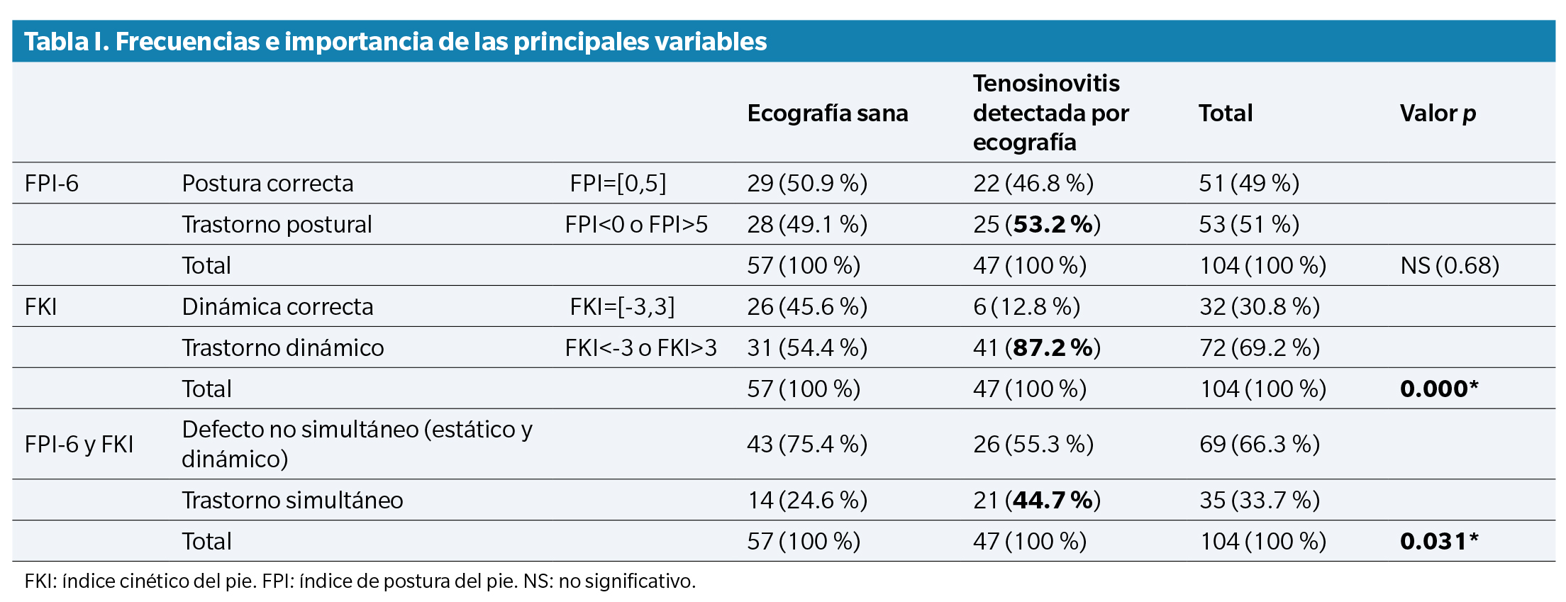
La relación observada entre el trastorno estático y la patología detectada en la ecografía no fue estadísticamente significativa (p = 0.68) y confirmó que solo 25 (53.2 %) de los 47 pies con patología detectada en la ecografía presentaron trastorno estático (FPI). Tampoco se observó significancia estadística (p = 0.89) en la relación existente entre la alteración estática y alteración dinámica del pie. También se analizó la asociación entre la patología detectada en la ecografía y el trastorno simultáneo, tanto estático como dinámico del pie, que también fue estadísticamente significativa (p = 0.031). De los 47 pies con tenosinovitis detectada por ultrasonido, 21 pies (44.7 %) presentaron trastorno simultáneo de FPI-6 y FKI (Tabla 1).
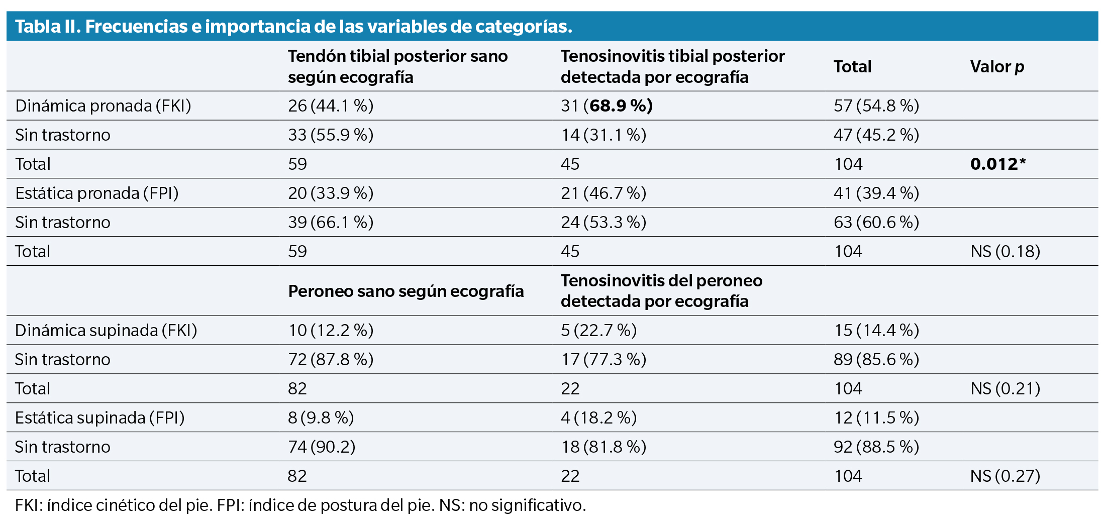
Se estudió la relación existente entre la tenosinovitis del tendón tibial posterior detectada por ecografía y el trastorno de pronación. Lo mismo ocurrió con la correspondencia entre la afectación de tendones peroneos y el trastorno de supinación. Los resultados mostraron que solo la queja de pronación dinámica se asoció con la tenosinovitis detectada del tendón tibial posterior por ecografía (p = 0.012). Otras relaciones no fueron estadísticamente significativas (Tabla 2).
Según el modelo de regresión logística binaria no ajustada empleado, se observó una tendencia significativa hacia una mayor probabilidad de tendinopatía del tobillo en participantes con marcha patológica. Las probabilidades no ajustadas de tendinopatía en pies con alteraciones dinámicas fueron OR = 5.9 (IC del 95 %: 2.15-16.26) frente a pies con marcha correcta (p = 0.001). Otros resultados de regresiones logísticas binarias, para trastornos estáticos o ambos trastornos en relación con la patología detectada por ecografía, no tuvieron significación estadística alguna.
Discusión
Este estudio mostró una asociación estadísticamente significativa entre los trastornos dinámicos y la tendinopatía del tobillo medida ecográficamente, un hallazgo que coincide con nuestra hipótesis de trabajo. El 87.2 % de los pies con trastornos dinámicos presentan tenosinovitis en la ecografía. Por su parte, el 44.7 % de los pies con ambos, trastornos estáticos y dinámicos, presentan tenosinovitis detectada en la ecografía. No obstante, los trastornos estáticos no presentan relación estadística alguna con la tendinopatía del tobillo, un nuevo abordaje biomecánico.
Los pies con trastorno dinámico podrían tener más probabilidades de presentar tendinopatías desde el punto de vista ecográfico. Esta podría ser la primera publicación sobre trastornos dinámicos como factor de riesgo para desarrollar tendinopatías. Algunos autores han buscado relaciones entre la postura estática y dinámica del pie y la biomecánica de la carrera, tal y como asegura la revisión realizada por Hollander(15). En cualquier caso, en la literatura médica existente, no hay antecedentes que nos impulsen a buscar relaciones entre estos dos trastornos (marcha patológica y tendinopatías del tobillo).
Nuestros hallazgos otorgan credibilidad al índice cinético del pie (FKI) para determinar trastornos dinámicos del pie, por lo que podría considerarse como una medida innovadora de la marcha. Será necesario desarrollar nuevos ensayos para evaluar la sensibilidad y especificidad del índice cinético del pie (FKI) como método diagnóstico. Las progresiones y características del COP se han estudiado en la literatura antes, pero su observación no ha demostrado ser capaz de diagnosticar trastornos dinámicos durante la marcha(16).
Las implicaciones de nuestros hallazgos son importantes por varias razones. En primer lugar, porque es necesario tener un valor cuantificable de los trastornos dinámicos tanto en el campo de la investigación como dentro de la práctica clínica destinada a determinar diagnósticos específicos. El índice cinético del pie nos ofrece precisamente eso. En segundo lugar, los resultados sugieren que tanto la toma de decisiones clínicas como el diseño de tratamientos se deben centrar en modificar los trastornos dinámicos. En tercer lugar, los trastornos dinámicos son un factor modificable que podría ser una diana para estrategias preventivas para atenuar las complicaciones de la tendinopatía, sobre todo en campos como la reumatología, la pediatría o la medicina deportiva. Controlar los factores de riesgo mejora los resultados de salud.
Nuestro estudio no está exento de limitaciones. Los hallazgos de las ecografías registrados en este estudio fueron asintomáticos, uno de los criterios de inclusión de los participantes fue la ausencia de dolor, sin síntomas. En este sentido, analizamos una etapa inflamatoria subclínica que en el campo del diagnóstico por ecografía ya se considera un trastorno patológico a pesar de no mostrar evidencia clínica evidente. Por otro lado, es necesario llevar a cabo un estudio de cohorte que confirmen los resultados de odds ratio obtenidos (OR = 5.9; IC del 95 %: 2.15,16.26) en nuestro trabajo, diseñado con carácter exploratorio, debido a la ausencia de antecedentes bibliográficos. Finalmente, la ausencia de un método de diagnóstico de la marcha dinámica validado, como gold estándar para poder realizar comparativas con el índice cinético del pie, nos llevó a diseñar este trabajo utilizando el método de diagnóstico por ecografía validado como sistema para comparar y ubicar asociaciones.
Nuestros datos vienen a reforzar varios abordajes biomecánicos nuevos e importantes. En primer lugar, no se observa que exista una relación entre una supinación estática o dinámica excesivas y patologías asociadas a los tendones peroneos, compartimento externo del tobillo. En segundo lugar, tampoco se observa una relación entre una pronación estática excesiva y la patología del tendón tibial posterior, compartimento interno del tobillo, aunque sí una relación entre una pronación dinámica excesiva y patologías del tendón tibial posterior detectada por ecografía. En tercer lugar, la asociación más alta (87.2 %, p = 0.000) se observa entre los trastornos dinámicos (en pronación, supinación o ambos) y la patología detectada por ecografía (para el tendón tibial posterior, los tendones peroneos o ambos). Los resultados sugieren que el control de los movimientos del tobillo en el eje sagital, pronación o supinación, no está regulado por un único grupo muscular tendinoso, del compartimento interno o externo del tobillo, respectivamente, sino que ambos son capaces de estabilizar los defectos de pronación/supinación. No obstante, los resultados vienen a confirmar que tal vez el que juega un papel más preponderante sea el tendón tibial posterior porque parece ser el que sufre más estrés o sobrecarga. Quizás por esta razón sea el tendón diana de lesiones de patología osteoartrítica.
En lo referente a las limitaciones y perspectivas de investigación de este artículo, destacamos que el índice cinético del pie (FKI) no es un índice validado. No pudimos encontrar ningún índice de evaluación validado ni ningún sistema diagnóstico de los trastornos dinámicos en la literatura científica. Existen criterios de clasificación patológica en función de los rangos de movilidad articular registrados por videocámaras de alta frecuencia, incluida la construcción de modelos en 3D para realizar valoraciones dinámicas. Consideramos, no obstante, excesivo el coste económico y la inversión de tiempo que estos sistemas de examen requieren, por lo que abogamos por la incorporación de un método de diagnóstico clínico asequible y extrapolable a patologías dinámicas, como el índice cinético del pie.
Confirmamos que los registros y evaluaciones fueron realizados exclusivamente por un único observador, para cada uno de ellos, por lo que la validez entre observadores no se pudo comparar. Debemos aclarar, no obstante, que este no fue el objetivo de nuestro estudio.
Consideramos que los modelos de regresión logística realizados en este estudio tienen la solidez necesaria para proporcionar datos iniciales que podrían ayudar a diseñar, basándose en ellos, otros estudios observacionales analíticos o cohortes o casos y controles, capaces de determinar los factores de riesgo y sus respectivos OR.
En conclusión, los trastornos de la función del pie durante la marcha están estadísticamente asociados a la tenosinovitis del tendón tibial posterior y/o peroneales detectada por ecografía. No se ha encontrado que los trastornos posturales del pie estén asociados a la patología tendinosa del tobillo detectada por ecografía. La tenosinovitis del tendón tibial posterior detectada por ecografía se asocia a la sobrepronación dinámica del pie y el tobillo durante la marcha. Los trastornos de la función del pie aumentan la probabilidad de tenosinovitis del tobillo detectada por ecografía. Una medida innovadora de la marcha, el índice cinético del pie, parece ser un puntaje diagnóstico capaz de detectar trastornos dinámicos. Esto requiere un proceso de validación, con diferentes sistemas de presión, un tamaño de muestra más grande y un diseño multicéntrico. Después de eso, el FKI podría emplearse como herramienta diagnóstica para facilitar la toma de decisiones clínicas y mejorar los tratamientos.
Agradecimientos
Agradecemos a Joan Teva su excelente trabajo, paciencia e interés desinteresados. Recopiló datos de evaluación estática de todos los participantes, siempre de forma rigurosa y eficiente; a la Dra. María Teresa Puig Reixach su disponibilidad y rigor científico en la realización de valoraciones críticas y mejora tanto del diseño como de la versión definitiva del presente estudio; a la Dra. Carmen Moragues su esfuerzo de tiempo en la recopilación inicial de muestras recogidas por ecografía.
Aprobación ética y consentimiento para participar
La aprobación ética para este estudio se solicitó al CEIC del “Institut de Recerca de l’Hospital de la Santa Creu i Sant Pau”, en Barcelona. Número de código: IIBSP-KPE-2017-48. Ref. HSCSP: 17/281 (OBS). Se solicitó autorización de la dirección del Departamento de Reumatología y se recopiló el consentimiento informado firmado de cada participante, antes de su aceptación formal como participante en el estudio, proporcionando una copia.
Financiación
Esta investigación no recibió ninguna subvención del sector público ni privado.
Declaraciones de interés
Ninguna.
Conflicto de intereses
Todos los autores declaran no tener ninguna relación financiera ni de otro tipo que pudiese llevar a un conflicto de intereses relacionado con este trabajo. Los autores son los únicos responsables del contenido y la redacción del manuscrito. Tampoco hay conflictos de intereses entre ninguno de los autores del presente artículo.
Disponibilidad de datos y materiales
El conjunto de datos utilizado y/o analizado durante el estudio actual está disponible gracias al autor de correspondencia bajo solicitud razonable.
Contribución de los autores
Concepción y diseño del estudio: ATC, CDT.
Recopilación de datos: ATC, PMA, FMS.
Análisis e interpretación de resultados: ATC, FMS.
Creación, redacción y preparación del borrador inicial del artículo: ATC.
Revisión y aceptación versión final previa publicación: ATC, CDT, PMA, FMS.
Bibliografía
- Zhu Y, Kang G, Yu C, Poh LH. Logarithmic rate based elasto-viscoplastic cyclic constitutive model for soft biological tissues. J Mech Behav Biomed Mater. 2016;61:397-409. DOI: 10.1016/j.jmbbm.2016.03.014.
- Criscenti G, De Maria C, Sebastiani E, Tei M, Placella G, Speziali A, et al. Quasi-linear viscoelastic properties of the human medial patello-femoral ligament. J Biomech. 2015;48(16):4297-302. DOI: 10.1016/j.jbiomech.2015.10.042.
- Thijs Y, De Clercq D, Roosen P, Witvrouw E. Gait-related intrinsic risk factors for patellofemoral pain in novice recreational runners. Br J Sports Med. 2008;42(6):466-71. DOI: 10.1136/bjsm.2008.046649.
- Sanchis-Sales E, Sancho-Bru JL, Roda-Sales A, Pascual-Huerta J. Dynamic Flexion Stiffness of Foot Joints During Walking. J Am Podiatr Med Assoc. 2016;106(1):37-46. DOI: 10.7547/14-141.
- Dowling GJ, Murley GS, Munteanu SE, Smith MM, Neal BS, Griffiths IB, et al. Dynamic foot function as a risk factor for lower limb overuse injury: a systematic review. J Foot Ankle Res. 2014;7(1):53. DOI: 10.1186/s13047-014-0053-6.
- Van der Leeden M, Steultjens M, Dekker JH, Prins AP, Dekker J. Forefoot joint damage, pain and disability in rheumatoid arthritis patients with foot complaints: the role of plantar pressure and gait characteristics. Rheumatology (Oxford). 2006;45(4):465-9. DOI: 10.1093/rheumatology/kei186.
- Rokkedal-Lausch T, Lykke M, Hansen MS, Nielsen RO. Normative values for the foot posture index between right and left foot: a descriptive study. Gait Posture. 2013;38(4):843-6. DOI: 10.1016/j.gaitpost.2013.04.006.
- Murley GS, Landorf KB, Menz HB, Bird AR. Effect of foot posture, foot orthoses and footwear on lower limb muscle activity during walking and running: a systematic review. Gait Posture. 2009;29(2):172-87. DOI: 10.1016/j.gaitpost.2008.08.015.
- Redmond AC, Crane YZ, Menz HB. Normative values for the Foot Posture Index. J Foot Ankle Res. 2008;1(1):6. DOI: 10.1186/1757-1146-1-6.
- Wong L, Hunt A, Burns J, Crosbie J. Effect of foot morphology on center-of-pressure excursion during barefoot walking. J Am Podiatr Med Assoc. 2008;98(2):112-7. DOI: 10.7547/0980112.
- De Cock A, Vanrenterghem J, Willems T, Witvrouw E, De Clercq D. The trajectory of the centre of pressure during barefoot running as a potential measure for foot function. Gait Posture. 2008;27(4):669-75. DOI: 10.1016/j.gaitpost.2007.08.013.
- Lugade V, Kaufman K. Center of pressure trajectory during gait: a comparison of four foot positions. Gait Posture. 2014;40(4):719-22. DOI: 10.1016/j.gaitpost.2014.07.001.
- Arnoldner MA, Gruber M, Syré S, Kristen KH, Trnka HJ, Kainberger F, et al. Imaging of posterior tibial tendon dysfunction--Comparison of high-resolution ultrasound and 3T MRI. Eur J Radiol. 2015;84(9):1777-81. DOI: 10.1016/j.ejrad.2015.05.021.
- Rabbito M, Pohl MB, Humble N, Ferber R. Biomechanical and clinical factors related to stage I posterior tibial tendon dysfunction. J Orthop Sports Phys Ther. 2011;41(10):776-84. DOI: 10.2519/jospt.2011.3545.
- Hollander K, Zech A, Rahlf AL, Orendurff MS, Stebbins J, Heidt C. The relationship between static and dynamic foot posture and running biomechanics: A systematic review and meta-analysis. Gait Posture. 2019;72:109-122. DOI: 10.1016/j.gaitpost.2019.05.031.
- Buldt AK, Forghany S, Landorf KB, Murley GS, Levinger P, Menz HB. Centre of pressure characteristics in normal, planus and cavus feet. J Foot Ankle Res. 2018;11:3. DOI: 10.1186/s13047-018-0245-6.